|
Featured Photos
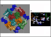 |
Location of modulatory beta subunits in BK potassium channels.
J. Gen. Physiol. 135:449-459; PMCID: PMC2860586.
Liu, G., Niu, X., Wu, R.S., Chudasama, N., Yao, Y., Jin, X., Weinberg, R., Zakharov, S.I., Motoike, H., Marx, S.O., and Karlin, A. (2010)
see Arthur Karlin, Ph.D. |
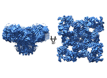 |
Ryanodine receptors channels are required for the release of calcium (Ca2+) from intracellular stores, a process that triggers cellular functions including excitation-contraction (EC) coupling in the skeletal and cardiac muscle. We solved a near-atomic resolution structure of this huge protein complex (2.3MDa), unraveling the structure of the full-length molecule and exposing new domains including the 6 transmembrane (TM) pore. see Andrew Marks Lab. |
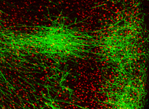 |
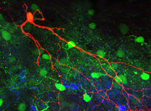
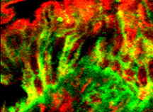
Callosal axon projections in a genetic mouse model of schizophrenia.
Axon terminal branching in the contralateral somatosensory cortex of EGFP-expressing neurons at 8 week-old mouse.
Green: EGFP, Red: NeuN. see Joseph A. Gogos, M.D., Ph.D.. |
 |
|
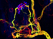 |
A branching mechanosensory neuron (red) that innervate skin cells (blue) integrates tactile information at nodes of Ranvier, which localize to gaps and endpoints of myelin (green). (courtesy of Kara L. Marshall, an Integrated graduate student in the Lumpkin laboratory) see Ellen Lumpkin Ph.D. |
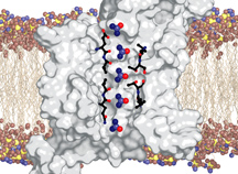 |
The urea transporter (UT) is responsible for rapidly transporting urea (a metabolic byproduct) across membranes. The transporter facilitates flow of urea (spheres) by forming a pore lined by conserved amino acids (sticks) . UT is necessary for the kidneys to concentrate urea and achieve water reabsorbtion. see Ming Zhou, Ph.D. |
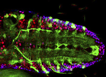 |
Circuits in the Drosophila Larval Ventral Nerve Cord. Just as in the human brain, the fruitfly Drosophila has complex arrangements of neurons interconnected by synapses to form circuits. The picture shows the Ventral Nerve Cord of a Drosophila larvae, a structure which shares some similarities of function to the human spinal cord. Motor neurons are in blue, interneurons are in red and a subset of motor neuron dendrites is labelled in green. see McCabe Lab |
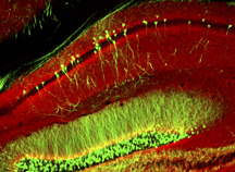 |
Hippocampus from GFP reporter mice counterstained with VGLUT1 (red),
a marker of excitatory synapses. Hypofunction of these synapses has
been proposed to be involved in the pathogenesis underlying several
psychiatric disorders. see Joseph A. Gogos, M.D., Ph.D. |
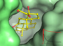 |
"Steroids modulate potassium channels"
Cortisone, a common drug treating inflammation, was found to interact with beta subunit of voltage-dependent potassium channels. A high resolution crystal structure of cortisone in complex with the beta subunit reveals a novel regulation site. The beta subunit is shown as surface representation in green, and cortisone is shown as ball-and-stick, with carbon in yellow and oxygen in red. Potassium currents recorded before (black) and after (red) cortisone application are show in the lower right corner. Ongoing research in Dr. Zhou’s lab is aimed at understanding the physiological functions and pharmacological regulations of potassium channels. see Ming Zhou, Ph.D. |
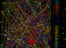 |
Connectivity patterns within a model neural network used to study how signals are propagated and routed from one region of the brain to another. Failure modes in this network are used to test theories of the role of interneurons in disorders such as schizophrenia and autism. see Larry Abbott, Ph.D. |
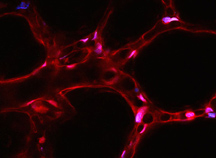 |
Live two-photon image of the lung shows microvascular network surrounding air-filled alveoli. Endothelial cells lining the vascular lumen show fluorescence in pseudocolor of the cytosolic dye, calcein (red) and the nuclear-staining dye, Hoechst 33342 (purple). The largest vessel is a venule (diameter, 20 microns) that collects blood from capillaries emanating from the inter-alveolar septum. Microvascular branches occurring normal to the image plane appear as circular structures. Such imaging is aimed at understanding the sites at which leukocytes cross the microvascular barrier and the routes they take to reach alveolar spaces. see Jahar Bhattacharya, M.D., Ph.D. |
 |
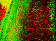
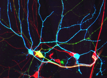
Location and morphology of inhibitory neurons in the dorsal horn are revealed by immunohistochemistry. The red cell is an example of a biocytin-filled EGFP-positive inhibitory neuron from a postnatal day 30 mouse. The cell body is at the lamina I/II border within the spinal cord, with processes extending as far ventrally as lamina III. The dense band of PCKg-positive somata and puncta (blue) reveals inner lamina II. Other EGPF-positive inhibitory neurons are also visible (green). Scale bars is 50 mm.
see Amy MacDermott, Ph.D. |
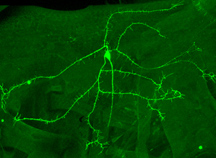 |
A neuron in the fruit fly Drosophila melanogaster labeled with a membrane-targeted green fluorescent protein. The cell body is in the center and is surrounded by exuberant dendritic processes. Ongoing research in the department is aimed at understanding how dendritic arbors develop their characterictic morphologies that are critical for proper nervous system function. see Grueber Lab |
 |
Expression of Hensin and Galectin 3 in the Colon. Hensin is a protein involved in terminal differentiation of epithelia. In the stem cell compartment (the crypts, right) hensin is only present in intracellular vesicles. In the terminally differentiated cells (the surface cells
(left) hensin is present in an extracellular pattern where it colocalizes with galectin 3, a protein necessary for its polymerization. see Qais Al-Awqati, M.D, CH.B. |
 |
Dendritic arbors of Drosophila sensory neurons showing self-avoidance behavior. Branches that arise from the same neuronal soma (cell bodies are near the bottom of the image) do not overlap, but branches of different cells can cross each other. Research in the department has shown that selective self-recognition and repulsion is mediated by the gene encoding Down syndrome cell adhesion molecule (Dscam). see Grueber Lab |
|
|