SNARE-mediated fusion at the single event level: the "flipped"SNARE cell-fusion assay
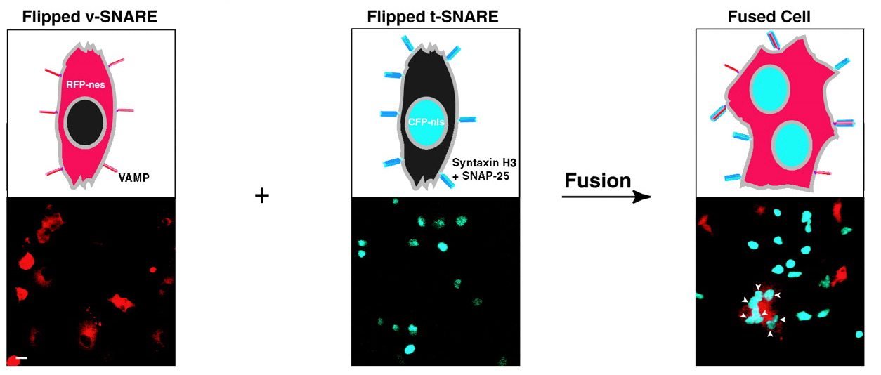
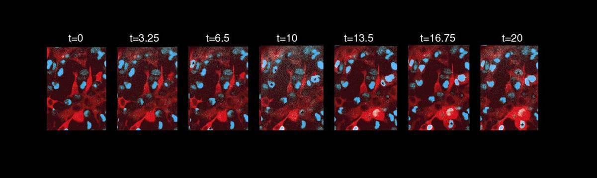
This assay also provided the first opportunity to visualize individual SNARE-mediated fusion events and to record them in real time (click on panels below for movie).
Membrane Fusion Intermediates
The identification of intermediate structures composed of both lipid and protein which may have physiologic lifetimes on the millisecond time scale is a daunting challenge. Our best understanding of how membranes merge comes from a large body of work on viral membrane fusion processes and is summed up in the stalk model of membrane fusion (roughly sketched on the left).
We are attempting to isolate intermediates on this pathway by manipulating the protein, lipid, and experimental conditions to trap membrane fusion at a particular step. For example, we have recently shown, with our flipped-SNARE cell-fusion assay (below), that when the transmembrane domain of a SNARE is replaced with a GPI anchor, membrane fusion is halted at a hemifusion stage, in which only the outer leaflets of the interacting cellular membranes mix. Whether we have isolated an on-pathway intermediate, or forced the membrane fusion reaction down a non-productive alternate pathway, is a question we continue to explore.